Biggest brain map ever details huge number of neurons and their activity

3D reconstruction is the first to overlay neuronal activity on a large-scale map of brain cells
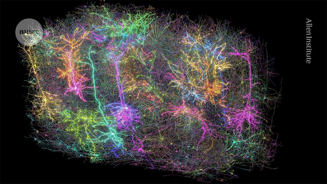
A rendering of more than 1,000 brain cells out of the those reconstructed from analysis of a cubic millimetre of brain tissue from a mouse. Credit: Allen Institute
Researchers have created the largest and most detailed wiring diagram of a mammalian brain to date, by mapping cells in a cubic millimetre of a mouse’s brain tissue1. In a landmark achievement, the diagram also details the activity of individual neurons on a large scale ― a neuroscience first.
The high-resolution 3D map contains more than 200,000 brain cells, around 82,000 of which are neurons. It also includes more than 500 million of the neuronal connection points called synapses and more than 4 kilometres of neuronal wiring, all found in a tiny block of tissue in a brain region involved in vision. The only brain map of comparable scale is that of a cubic millimetre of human brain, which included 16,000 neurons and 150 million synapses2. The new map also captured the activity of tens of thousands of neurons firing signals and interacting with each other to process visual information.
This brain-activity map, combined with the wiring diagram, marks a milestone in connectomics, a field that aims to show how brains process and organize information. Behind the massive efforts are more than 150 researchers in the Machine Intelligence from Cortical Networks (MICrONS) project, who described their work in a package of eight papers published today in Nature and Nature Methods. The MICrONS project has made its resources available for the neuroscience community online, and other teams are already exploring them in different studies.
“They managed to do something that we haven’t done as a neuroscience community in basically all of our history, which is to be able to map the activity of neurons onto the wiring on a very large population of neurons,” says Mariela Petkova, a neuroscientist at Harvard University in Cambridge, Massachusetts, who is not involved with the project. “We have never seen it at this scale.”
The data “are really stunningly beautiful,” says Forrest Collman, a neuroscientist at the Allen Institute for Brain Science in Seattle, Washington, who co-authored the studies. “Looking at it really gives you an awe about the sense of complexity in the brain that is very much akin to looking up at the stars of night.”
Mouse in a matrix
To create the breakthrough map, researchers first recorded the firing of almost 76,000 neurons in the visual cortex of a mouse as the animal watched various videos, including clips from The Matrix, for two hours. Then they sliced up a cubic millimetre of the mouse’s brain into thousands of tissue slices, each about one four-hundredth the width of a human hair.
The scientists imaged each slice and assembled the images into a 3D map. Finally, they used artificial intelligence and machine-learning algorithms to annotate the neurons, their branching projections and their synapses. The team also matched the neurons in the map with their recordings of brain cells in action.
Moritz Helmstaedter, a neuroscientist at the Max Planck Institute for Brain Research in Frankfurt, Germany, says “the combination of function and structure at that scale” is unprecedented. It’s “a very impressive endeavour and success”.
Fire together, wire together
Enjoying our latest content?
Login or create an account to continue
- Access the most recent journalism from Nature's award-winning team
- Explore the latest features & opinion covering groundbreaking research
or
Sign in or create an account Continue with Google
 Continue with ORCiD
doi: https://doi.org/10.1038/d41586-025-01088-x
Read the related News & Views, ‘A vast brain map links neural activity and wiring’.
This story originally appeared on: Nature - Author:Miryam Naddaf


