How to keep the lights on: the mission to make more photostable fluorophores

Fluorescent labels that have greater resistance to bleaching could help researchers to get more from biological imaging
Fluorescence microscopy is one of the most powerful tools in the life-sciences toolkit. The unique spectral properties of fluorescent dyes and proteins, collectively called fluorophores, have allowed scientists to capture snapshots — and even movies — of the microscopic universe that makes biology tick. Yet scientists are still perplexed by the fundamental chemistry that keeps the lights on.
All fluorophores work on the same principle: energy absorbed from an incoming photon excites the molecule to a higher energetic state. As the fluorophore ‘relaxes’, it dissipates energy by emitting a new photon of a different (longer) wavelength. The more efficiently a fluorophore absorbs photons and radiates light, the stronger its fluorescent signal.
The problem is that fluorophores can’t perform this light show forever: they dim over time in a phenomenon called photobleaching. Photobleaching occurs more rapidly with high-intensity light, and after repeated rounds of excitation and in oxygen-rich environments. This can end experiments prematurely and sometimes generates toxic by-products that can kill cells. Some of the earliest dyes would fade in seconds under bright light, whereas many of today’s fluorophores can stay bright for minutes or more under the right conditions.
Photobleaching has become an irritating puzzle in the world of biological imaging, but it’s neither glamorous nor easy to solve. Much remains unknown about the chemistry that drives or prevents it, says Luke Lavis, an organic chemist at the Howard Hughes Medical Institute’s Janelia Research Campus in Ashburn, Virginia. Strategies that fortify a fluorophore against bleaching might inadvertently affect other spectral characteristics. And changes that improve one fluorophore can be difficult to apply to others. “It’s the sort of thing that people just live with rather than really trying to study,” says Adam Cohen, a neuroscientist at Harvard University in Cambridge, Massachusetts.
Turning on the lights
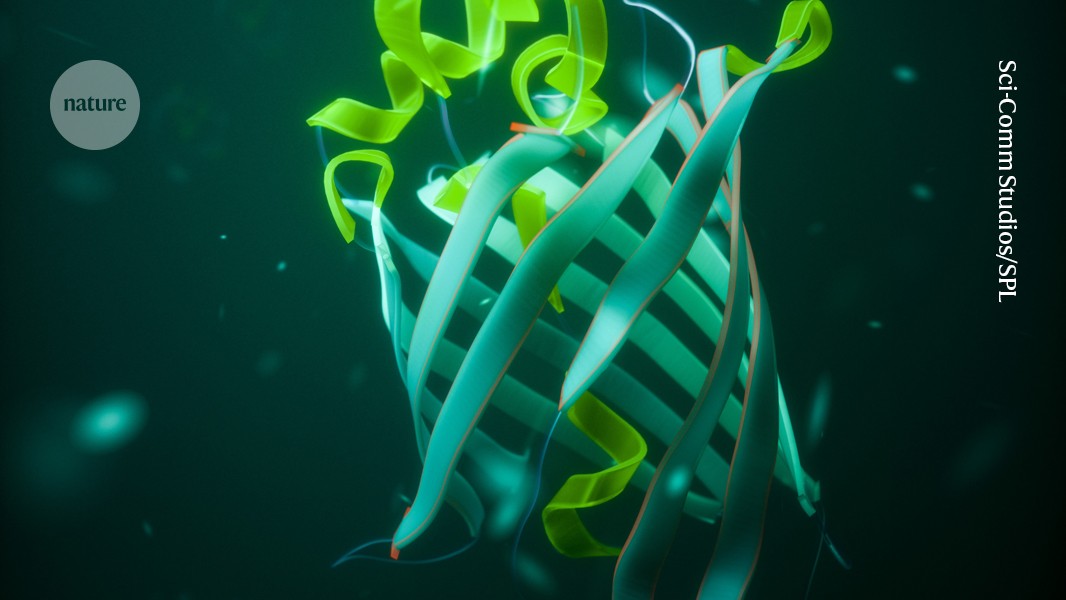
Microscopists have a vast palette of dyes and fluorescent proteins at their disposal. Microscope vendor Leica Microsystems, based in Wetzlar, Germany, lists more than 250 fluorescent dyes for life-sciences imaging, and the FPbase fluorescent protein database includes nearly 1,100 entries. Fluorescent dyes are descendants of the nineteenth-century dye industry, when chemists were first learning to make small organic molecules such as fluorescein, rhodamine and cyanine dyes. Fluorescent proteins stem from the discovery in 1962 that certain jellyfish fluoresce because they contain a family of proteins in which a coloured molecule called a chromophore, built from amino acids, is encased in a protective barrel formed from a type of protein structure called β-pleated sheets (corals were subsequently also found to contain the same protein family).
Whatever their chemical nature, these molecules are driven by the same photophysical principles. When a molecule is illuminated by incoming light, the energy from the absorbed photon excites electrons in the material. The fluorophore can’t hold on to that energy forever — the time it spends in the excited state is called the fluorescence lifetime. Eventually it must relax, ideally by offloading that pent-up energy as radiated photons: one photon is released for every photon absorbed.
In practice, excited electrons find myriad non-radiative pathways to return to their ground state — their normal energy level — without emitting a photon. The fluorophore might lose energy through collisions with other molecules or through vibrations in its own bonds, for instance. Off-target photoreactions might generate toxic reactive oxygen species or even destroy the fluorophore. In both cases, fluorescence is temporarily or permanently snuffed out, and quantum yield — the ratio of fluorescence out to excitation light in — suffers.
But it’s rarely clear why that is. Scientists have struggled to isolate bleached fluorophores to do a chemical post-mortem, and it’s nearly impossible to predict all of the non-radiative pathways with which fluorescence competes. But the inevitability of photobleaching means that microscopists have only a limited ‘photon budget’ with which to work, and they must spend it carefully before the fluorophore — and their experiment — goes dark.
One way to protect that return on investment is by shutting down the competition. Researchers have long noted a correlation between a fluorophore’s brightness and the flexibility of its chemical backbone, says Ralph Jimenez, a physical chemist at the University of Colorado Boulder. Stiff molecules tend to have longer fluorescence lifetimes, which corresponds to higher quantum yield, he says.
“A lot of synthetic chemists tried to make fluorophores brighter by making them very rigid so that basically nothing could wiggle any more,” says chemical biologist Michelle Frei at the Swiss Federal Institute of Technology in Zurich. The drawback was that these ‘rigidified’ molecules were more likely to get trapped in cell membranes before reaching their target, making them less useful in cellular assays, Frei says.
Rigidification has benefits for fluorescent proteins, too, says Dorus Gadella, a molecular scientist at the University of Amsterdam. The chromophore — which is highly conserved across fluorescent proteins — consists of two rings bridged by a methylene group. Yet this molecule does not fluoresce when excited in isolation, Gadella says, because it undergoes a molecular rearrangement known as cis–trans isomerization — a non-radiative pathway that diverts energy from emitting photons. The protein’s β-barrel rigidifies the chromophore and prevents that rearrangement from happening, he says.
Better fluorescent proteins
Working with biochemist Amy Palmer, also at the University of Colorado Boulder, Jimenez and his colleagues developed a bright red fluorescent protein called mCherry-XL, a variant of the popular mCherry. They did so by rigidifying the protein barrel and locking down a squiggly amino-acid side chain that was hampering fluorescence1; this helped to increase the fluorescence lifetime from 1.6 nanoseconds for mCherry to 3.9 nanoseconds for mCherry-XL. The trade-off is that the more time a fluorophore spends in its excited state, the more likely it is to become damaged, Jimenez says. As a result, mCherry-XL is three times brighter than mCherry, but less photostable.
Too much rigidity, however, can interfere with the protein’s ability to build its protective barrel and perform the multi-step, oxygen-dependent reaction that forms its chromophore, which can slow down the protein’s maturation or quash its fluorescence. A red fluorescent protein developed by Gadella and his colleagues, called mScarlet3, nicely illustrates this balancing act.
Cell nuclei in a zebrafish larva labelled with the red fluorescent protein mScarlet3.Credit: T. W. J. Gadella Jr et al./Nature Methods
According to Gadella, their protein represents a sweet spot in stiffening the barrel so that the chromophore can’t isomerize, but still leaves enough wiggle room for the protein to fold and mature. Thanks in part to its fast maturation rate, mScarlet3 is the brightest red fluorescent protein so far — more than five times brighter than mCherry — and it has a fluorescence lifetime of 4 nanoseconds2. But, much like mCherry-XL, mScarlet3 photobleaches faster than its precursors do under intense illumination.
Theoretical chemist Anna Krylov at the University of Southern California in Los Angeles, is developing software that can simulate excited-state processes in fluorophores to enhance their fluorescent properties. By identifying and shutting down non-radiative pathways, the thinking goes, fluorophores can be improved so they are fit for experimental purposes.
Researchers have observed, for instance, that photo-oxidation of the green fluorescent protein EGFP can turn its output from green to red. This behaviour is not observed in the closely related yellow fluorescent protein EYFP, despite both proteins having the same chromophore structure. Using their simulations, in 2016 Krylov and her team uncovered a non-radiative pathway in EGFP through which an electron can hop from the chromophore to a tyrosine residue in the protein barrel and thence to an electron acceptor outside the protein molecule3. That electron-transfer mechanism alters the photophysical properties of the EGFP chromophore, turning it red. In EYFP, however, the comparable tyrosine is chemically out of reach from the chromophore, preventing that electron transfer.
The calculations suggested that the team could suppress the non-radiative pathway in EGFP by swapping the tyrosine for leucine, which lacks tyrosine’s electron-hungry oxygen atom. “We were able to propose specific mutations to our experimental collaborators and were able to get a more photostable variant of this protein,” Krylov says. In fact, the resulting mutant was 80 times more photostable than EGFP, but was also dimmer. Although this mutant protein is not suitable for use in biological imaging, it demonstrates how a better understanding of fluorophore photophysics can lead to rational engineering of bespoke materials, Krylov says.
That said, serendipity and random mutagenesis still play an outsized part in the development of fluorescent proteins, Jimenez says. Atsushi Miyawaki, a biochemist at the RIKEN Center for Brain Science near Tokyo, and his team developed what could be the most photostable fluorescent protein so far. They discovered a remarkably photostable, but dim, green fluorescent protein in Cytaeis uchidae, a small jellyfish found along the Japanese coast.
Through random mutagenesis, the researchers found that they could improve the protein’s brightness without compromising photostability by changing a single amino acid from valine to alanine, which increased the protein’s folding and maturation efficiency4. Called StayGold, this mutant has a nearly identical structure to the original protein, yet emits ten times more photons before photobleaching than any previous green fluorescent protein, allowing the team to record the dynamic rearrangement of intricate organelle networks in live cells for up to six minutes5.
New chemistry
Fluorescent proteins and dyes each have their advantages. Proteins can be expressed in specific cell types, for instance, whereas dyes, as small molecules, are brighter and more easily fine-tuned through synthesis. Now, researchers have developed a fluorophore system that combines the best of both, according to Frei. Self-labelling proteins, such as the commercially available SNAP-tag and HaloTag reagents, work by embedding an enzymatic protein tag in a target of interest. This tagged protein can then be coupled to any of several small fluorescent molecules supplied by the researcher. For Frei, this design provides a platform for fluorescence lifetime imaging (FLIM), a photon-intensive method that differentiates fluorophores by how long they spend in the excited state rather than by the wavelength they emit.
As a doctoral student at the Max Planck Institute for Medical Research in Heidelberg, Germany, Frei created a panel of HaloTag variants that included mutations in the dye docking site. Mutations that helped to hold the fluorophore in place, she saw, extended fluorescence lifetimes — even though the fluorescent dye molecule itself was unchanged. Exploiting this finding, Frei engineered cells to express different self-labelling HaloTag proteins, so they could be treated with the same rhodamine dye but would elicit measurably different signals for each target6. Whereas conventional fluorescence microscopy experiments can handle only one dye per colour channel, Frei and her team were able to visualize three targets in a single-channel experiment by adding FLIM to the mix.

Fibroblast cells labelled with three fluorescent colours: blue for nuclei, green for microtubules and red for cell–cell contacts.Credit: Dr Jan Schmoranzer/Science Photo Library
To take full advantage of such self-labelling systems, however, fluorescent dyes would need an upgrade. Dye chemistry has changed little since the nineteenth century and requires harsh conditions that are ill-suited to building highly tailored molecules. In a 2023 review, Lavis and Martin Schnermann at the US National Cancer Institute in Frederick, Maryland, argued that chemists could rejuvenate classic dye scaffolds for next-generation microscopy by applying modern synthetic techniques7.
Take the classic red dye tetramethylrhodamine (TMR), for example. Lavis and his colleagues reasoned that rotation of the bond between its carbon skeleton and a structure called dimethylamine in the excited state might drive electron transfer to a non-radiative pathway. Chemists have conventionally suppressed that pathway by making rhodamine dyes in which these nitrogen-containing groups are locked in place with clunky fused-ring systems, which improved brightness but hindered permeability when the dye was applied to cells. Then, in 2015, Lavis and his collaborators demonstrated8 that a palladium-catalysed reaction could be used to install nitrogen-containing rings of different sizes in place of the original dimethylamine. Lavis’s team used these comparatively sleek functional groups to block rotation of the troublesome bond without the permeability challenges of previous analogues. One such molecule, a TMR analogue with a four-membered ring, had double the quantum yield of its parent, the researchers found.
But that’s in isolation. The team incorporated the molecule, named Janelia Fluor 549, into a self-labelling HaloTag system for cell imaging. When the researchers expressed the chromosomal proteins called histones in live cultured cells labelled with the Janelia Fluor 549 HaloTag ligand, they emitted nearly twice as many photons per second as the commercially available TMR-based HaloTag ligand — and for twice as long8. Furthermore, swapping out the dimethylamine groups on other scaffolds — including rhodamines and coumarins — yielded similar improvements to brightness without noticeable side effects.
Photostability in action
Lavis and his colleagues have now expanded the Janelia Fluor series to include a variety of functional groups, such as fluorinated and deuterated substituents9, and the dyes are already enabling discoveries. For example, Cohen’s lab at Harvard University uses high-resolution fluorescence microscopy to watch voltage changes as individual neurons fire in the brains of living mice. “It’s a very noisy environment,” Cohen says. “You need to be able to pick out these tiny fluctuations in fluorescence that only last a millisecond.” That means Cohen needs to illuminate his cells at a high frame rate to capture signal transduction in action — a photon-intensive experiment and a classic formula for photobleaching. “If you’re trying to get a lot of photons out of your molecules in a very short time, you need to drive them hard — you need to put a lot of light onto them — and that means they have to be photostable,” Cohen says.
In the past, researchers have studied signal transduction in neurons mostly on the basis of voltage changes at the cell body. Neuronal cell bodies receive inputs from dendrites that are ten times thinner than the bodies and are long enough for the voltage difference at the tip of a dendrite to be different from that at its base. It has not been possible to observe these differences using existing fluorescence systems, Cohen says. By better illuminating how electrical signals propagate through dendrites, photostable fluorophores could reveal how neurons process information and learn.
Cohen, Lavis and their colleagues have now combined the red-shifted dye Janelia Fluor JFX608 with a self-labelling, genetically encoded voltage-indicator system to record sub-millisecond voltage changes across dendritic trees in individual neurons in slices of mouse brain10. In a second study using the same fluorophore system, the researchers observed voltage changes in the neurons of live mice as they responded to external stimuli11. Both papers were first posted on the bioRxiv preprint server in 2023 and are undergoing peer review.
Still, what works for one dye doesn’t necessarily apply to others, and it’s notoriously difficult to compare one fluorophore to another. The key, Gadella suggests, is to get into the lab: users need to test different fluorophores to make sure their molecule is fit for purpose. Like many researchers, Gadella and his team make their materials available through the non-profit organization Addgene, which has already processed more than 700 orders for mScarlet3. Lavis says his lab has shared some 20,000 aliquots of Janelia Fluor dyes through Janelia’s Open Science program.
More dyes and more photons — that’s a recipe that Lavis, Gadella and others hope will allow researchers to observe complex biochemical phenomena that have so far escaped our view.
Nature 630, 258-260 (2024)
doi: https://doi.org/10.1038/d41586-024-01591-7
This story originally appeared on: Nature - Author:Ariana Remmel


