How the brain spies on the gut: with help from newfound immune cells

The body’s pathogen fighters can act as surveillance forces that ferry information from the gut and fat deposits to the brain
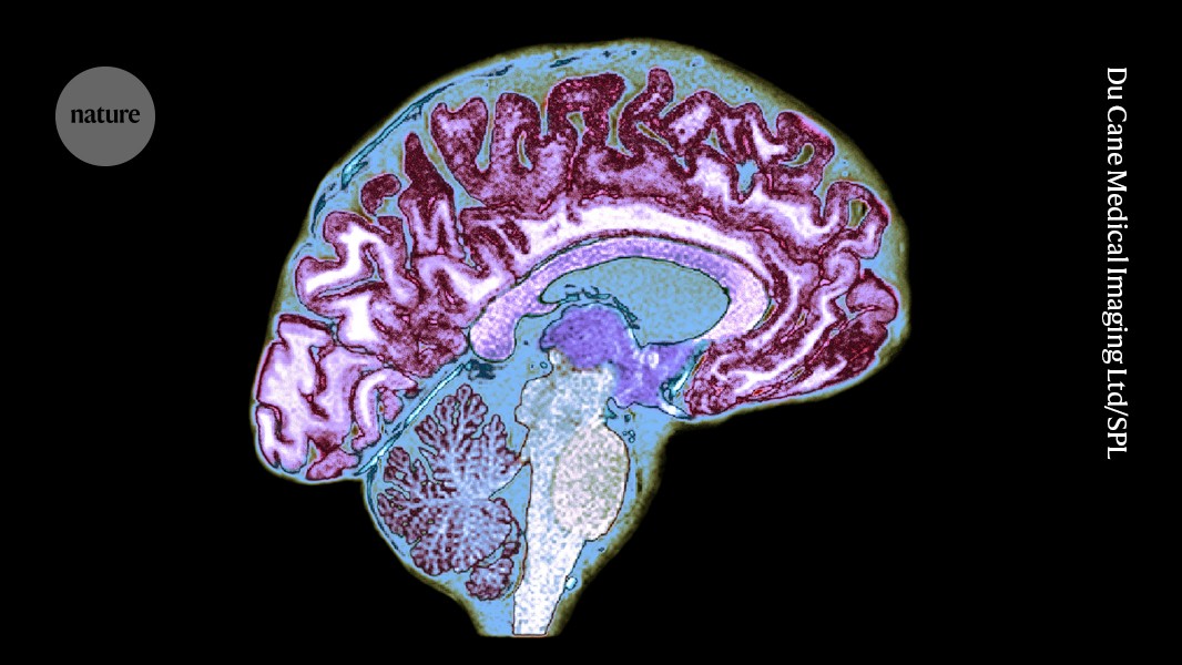
An area buried deep in the brain is a hotspot for immune cells called T cells.Credit: Du Cane Medical Imaging Ltd/Science Photo Library
Special-agent immune cells carry information about the gut and fat tissue deep into the brain. This surveillance system, uncovered in mice, is crucial to the brain’s control of behaviour such as pursuit of food, researchers report today in Nature1.
Similar immune cells are known to populate the membranes covering the brain, but the newly identified cells have molecular traits that give them entry into the core of the organ. Their function is shaped by diet and the microbiome; without them, even hungry mice are slow to eat.
“It’s very exciting to see that from an organ that was considered completely isolated from the immune system,” says Michal Schwartz, a neuroimmunologist at the Weizmann Institute of Science in Rehovot, Israel, who was not involved in the study.
Neighbourhood watch
Almost every organ in the body houses its own immune cells. Researchers have long thought that most of the healthy brain’s ‘adaptive’ immune cells — those that target specific pathogens — reside in the meninges, the three protective membranes enclosing the brain.
Previous studies have hinted that adaptive immune cells called T cells might dwell inside the brain itself under stable conditions. But determining whether these cells are truly distinct from those in the meninges has been a challenge, says Tomomi Yoshida, a neuroimmunologist at Yale University in New Haven, Connecticut and a co-author of the latest work. “They didn’t really compare and contrast that element,” she says.
To gain a more definitive answer, Yoshida and her colleagues spent five years searching for T cells across the entire mouse brain. They found that T cells are concentrated in a hotspot in the subfornical organ, a structure in the centre of the brain that regulates a range of bodily processes, including eating and drinking. The scientists then sampled tissue from the human subfornical organ, where they also found T cells.
In both mouse and human brains, the subfornical T cells were distinct from those found in the meninges. They produced more proteins that enabled them to reside in brain tissue and secreted more immune-signalling proteins known as cytokines under normal conditions.
Curiously, the researchers found that the T cells from inside mouse brains were very similar to those in the animals’ fat deposits. To understand this similarity, they gave some mice a high-fat diet. These mice ended up with more T cells in their fat and brain tissues than did mice that ate standard food — evidence of a link between fat mass and brain T-cell populations. After mice on the same diet fasted for 48 hours, the number of T cells in their brains rose and the number in their fat fell, suggesting that food intake affects the number of T cells travelling to the brain.
Enjoying our latest content?
Login or create an account to continue
- Access the most recent journalism from Nature's award-winning team
- Explore the latest features & opinion covering groundbreaking research
or
Sign in or create an account Continue with Google
 Continue with ORCiD
doi: https://doi.org/10.1038/d41586-025-01655-2
This story originally appeared on: Nature - Author:Gemma Conroy


