Mini hearts, lungs and livers made in lab now grow their own blood vessels

These sophisticated models will be used for human-development studies and drug testing
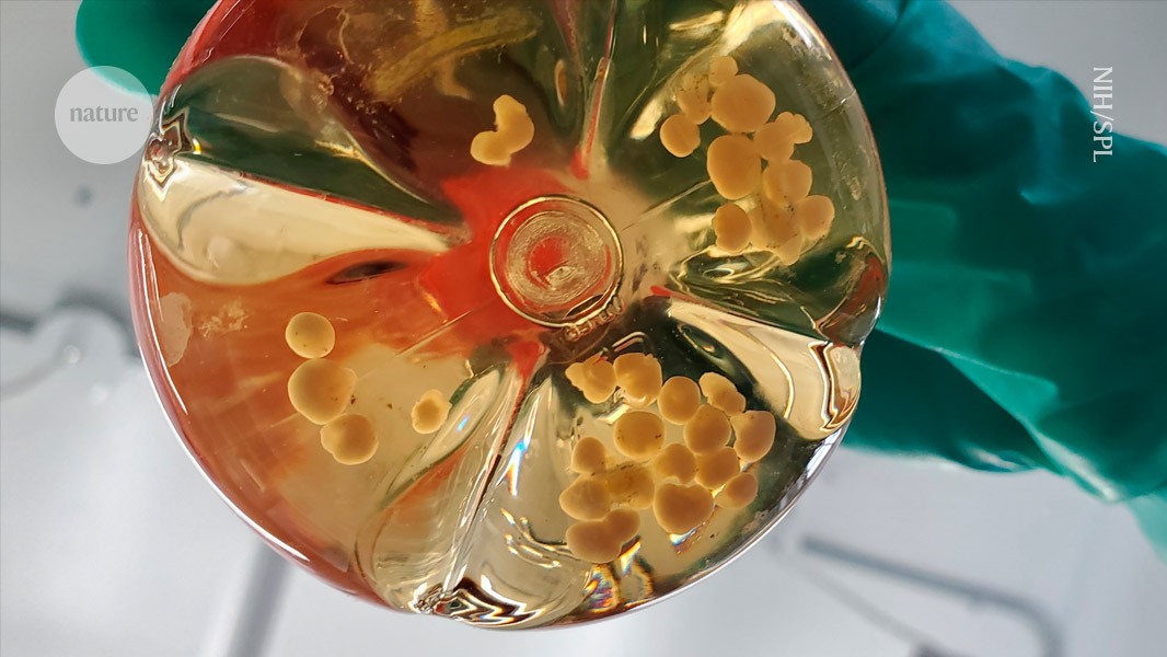
Human cerebral organoids inside a flask. Scientists have grown hearts, liver and lung organoids with blood vessels.Credit: National Institutes of Health/Science Photo Library
Researchers are making ever more sophisticated mini organs in the lab — and now they can grow their own blood vessels. The structures, which mimic the heart, liver, lungs and gut, are some of the most complex models of human development ever made, and contain cell populations and structures not seen before in these models.
“Vascularization of organoids is a hot topic,” says Ryuji Morizane, a nephrologist and stem-cell biologist at Massachusetts General Hospital in Boston.
Miniature 3D cell structures, called organoids, have been used for many years to test drugs and study disease and development. But most organoids lack the vessels that transport blood, nutrients and oxygen throughout the body, which has restricted their size, function and ability to mature. Kidneys, for example, need vessels to filter blood and produce urine, and lungs need them to exchange oxygen and carbon dioxide.
Last month in Science1 and Cell2, two separate teams reported creating vascularized organoids using a new approach that grows the organoids with vessels from their earliest stages. Starting with pluripotent stem cells, which can transform into almost any cell type in the body, the researchers coaxed the cells to form vessels as they were making the other organ tissue.
The models “really show the power of this strategy”, says Oscar Abilez, a stem-cell biologist at Stanford University in California, who co-authored the heart and liver paper1.
Early work
The first attempts to vascularize organoids involved growing blood vessel tissue in a dish and then combining it with other cell types to form ‘assembloids’3. But these models were still limited in their ability to mimic the structure and maturity of real organs.
Researchers stumbled on the new approach by accident. While growing epithelial cells, which form the outer lining of some organs and tissues, several research teams, including a group at the University of Michigan, Ann Arbor, reported4 that their organoids also spontaneously formed non-epithelial cells of a type that lines the blood vessels. These cells are typically seen as ‘contaminants’ to get rid of, but the Ann Arbor group tried to amplify that contamination in intestinal organoids.
Capitalizing on the surprise find by other groups, Yifei Miao, a stem-cell biologist at the Institute of Zoology, Chinese Academy of Sciences, in Beijing and his colleagues decided to see whether they could control the simultaneous growth of both types of cell — epithelial and blood-vessel cells — in the same dish, starting with lung and gut organoids.
But in early development, epithelial and blood-vessel cells require entirely opposing molecular triggers to grow. “You can either save one or the other one, so naturally they couldn’t grow together,” says Miao. He and his colleagues found a way to time the dosing of a cocktail of molecules to trigger the formation of both types of tissue at once from stem cells2.
Login or create a free account to read this content
Gain free access to this article, as well as selected content from this journal and more on nature.com
or
Sign in or create an account Continue with Google
 Continue with ORCiD
doi: https://doi.org/10.1038/d41586-025-02183-9
This story originally appeared on: Nature - Author:Smriti Mallapaty


